Scientists from Spain have recently compared the efficacy of the quantitative polymerase chain reaction (qPCR) method and immunohistochemistry in detecting severe acute respiratory syndrome coronavirus 2 (SARS-CoV-2) in different human tissues.
They have found that qPCR is a significantly more effective and sensitive method than immunohistochemistry in detecting SARS-CoV-2 in both cytology and formalin-fixed paraffin-embedded tissue samples.
The study is currently available on the Research Square* preprint server while under consideration at Scientific Reports.
Background
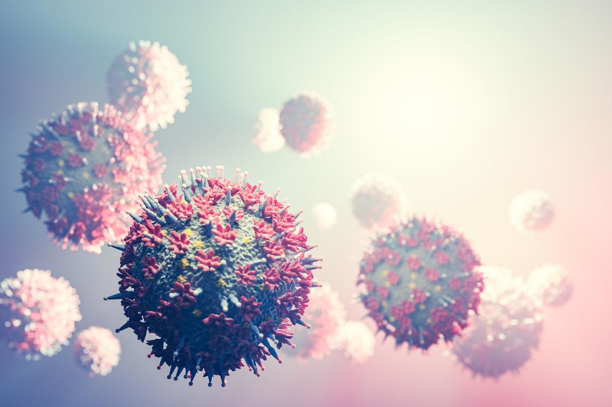
Severe acute respiratory syndrome coronavirus 2 (SARS-CoV-2), the causative pathogen of coronavirus disease 2019 (COVID-19), is an enveloped, positive-sense, single-stranded RNA virus belonging to the human beta-coronavirus family. The clinical diagnosis of COVID-19 is primarily done by qPCR method using nasopharyngeal (respiratory) samples. In addition, serological detection of anti-SARS-CoV-2 antibodies plays a vital role in detecting both acute and previous infections.
Being a respiratory virus, SARS-CoV-2 primarily infects epithelial cells present in the upper and lower respiratory tracts. However, the presence of the virus has also been detected in other tissues, including intestinal, cardiac, hepatic, brain, and kidney tissues. Despite the multiorgan involvement in COVID-19, only a limited number of studies are available to describe the detection of SARS-CoV-2 in different human tissues through immunohistochemistry. The available literature suggests the simultaneous use of two antibodies targeting the viral nucleocapsid and spike protein for immunohistochemical detection of the virus.
Besides the application of direct immunofluorescence and in situ hybridization methods for detecting SARS-CoV-2 in paraffin tissue sections, qPCR method has also been used to detect the virus in formalin-fixed paraffin-embedded samples of tongue squamous cell carcinoma.
In the current study, the scientists have validated and compared the efficiency of immunohistochemical and qPCR methods in detecting SARS-CoV-2 in both cytology and formalin-fixed paraffin-embedded tissue samples.
Important observations
A wide-variety of samples was used for the analysis, including small biopsy, surgical, liquid cytology, autopsy, and cytology block samples. These samples were mostly collected from the kidney, lung, placenta, and esophagus.
A total of 43 cytology and formalin-fixed paraffin-embedded tissue samples were included in the analysis. These samples were collected from patients who had a history of qPCR-confirmed SARS-CoV-2 infection.
Three relevant time periods were estimated in the study, including the interval time, SARS-CoV-2 persistence time, and archival time. The interval time defines the duration between the first qPCR-confirmed nasopharyngeal positive result and the collection of tissue/cytology samples. The persistence time defines the duration between the first qPCR-confirmed nasopharyngeal positive result and the subsequent first qPCR-confirmed nasopharyngeal negative result. The archival time defines the duration between sample collection and RNA extraction.
The estimation of these time periods revealed that the average interval time, persistence time, and archival time were 34 days, 28 days, and 220 days, respectively.
Quantitative polymerase chain reaction
The analysis of patient-derived cytology and formalin-fixed paraffin-embedded tissue samples by qPCR revealed SARS-CoV-2 positivity in about 25% of the samples. The formalin-fixed paraffin-embedded tissue samples included for the analysis were collected from the kidney, esophagus, lung, and placenta. In addition, ascitic fluid, pleural fluid, and urine were used as cytological samples.
Immunohistochemistry
A robust immunohistochemistry positivity for both anti-spike and anti-nucleocapsid antibodies was observed only in one placental sample with acute villitis. In addition, weak immunohistochemical positivity signals were detected in four samples, including pneumocytes in the lung, microglia in the brain, tubular epithelial cells in the kidney, and inflammatory cells in the placenta.
All immunohistochemistry-positive samples also showed positivity in the qPCR method. The only sample with strong immunohistochemical signal corresponded with the lowest cycle threshold (Ct) value of the qPCR method. A low Ct value in PCR represents high viral load.
Association between viral load and time periods
The interval, persistence, and archival time periods were correlated with qPCR-confirmed viral load. The analysis revealed a significant correlation of viral load with interval time, virus persistence time, and archival time.
Overall, these observations indicate that shorter interval and persistence time is associated with higher viral load in the samples.
Study significance
The study reveals that qPCR can be used to detect SARS-CoV-2 in both cytology and formalin-fixed paraffin-embedded tissue samples and that the method is more sensitive and accurate than immunohistochemistry.
*Important notice
Research Square publishes preliminary scientific reports that are not peer-reviewed and, therefore, should not be regarded as conclusive, guide clinical practice/health-related behavior, or treated as established information.
- Bernal-Florindo I. 2022. Identification of SARS-CoV-2 in different human tissues. Validation of immunohistochemical and qPCR techniques in paraffin-embedded tissues and cytology. Research Square. doi: 10.21203/rs.3.rs-1441884/v1 https://www.researchsquare.com/article/rs-1441884/v1
Posted in: Medical Science News | Medical Condition News | Disease/Infection News
Tags: Antibodies, Biopsy, Brain, Carcinoma, Cell, Coronavirus, Coronavirus Disease COVID-19, covid-19, CT, Cytology, Efficacy, Hybridization, Immunohistochemistry, Kidney, Microglia, Nasopharyngeal, Pathogen, Placenta, Polymerase, Polymerase Chain Reaction, Protein, Research, Respiratory, Respiratory Virus, RNA, RNA Extraction, SARS, SARS-CoV-2, Severe Acute Respiratory, Severe Acute Respiratory Syndrome, Spike Protein, Squamous Cell Carcinoma, Syndrome, Tongue, Virus

Written by
Dr. Sanchari Sinha Dutta
Dr. Sanchari Sinha Dutta is a science communicator who believes in spreading the power of science in every corner of the world. She has a Bachelor of Science (B.Sc.) degree and a Master's of Science (M.Sc.) in biology and human physiology. Following her Master's degree, Sanchari went on to study a Ph.D. in human physiology. She has authored more than 10 original research articles, all of which have been published in world renowned international journals.
Source: Read Full Article


