What are phosphors and how are they used?
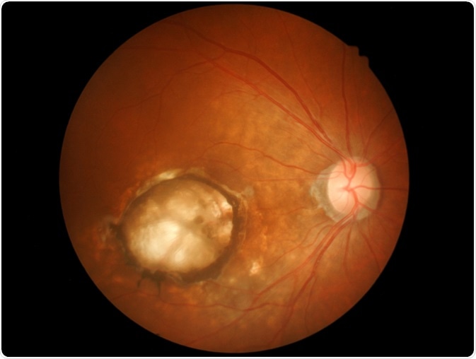
Image Credit: Memorisz/Shutterstock.com
Phosphors refer to a group of solid materials that emit visible light, or luminesces, in response to exposure to radiation in the form of ultraviolet light or an electron beam. Mostly, phosphor nanoparticles do not initiate a response or interaction when introduced to biological tissue, meaning that they are biologically inert.
Therefore, they can be used to study cells and biological substances as they can be injected into cells or used as probes when added to antibodies. Phosphors that are fluorescent under ultraviolet light or multiphoton excitation are suitable for use as markers in correlative microscopy.
Currently, the number of phosphors that have been successfully synthesized goes into the hundreds of thousands, and each of these has its characteristic color and emission period.
There are a wide number of applications of phosphors, those that fluoresce from electron excitation, known as electroluminescence, are used in the production of cathode-ray tube (CRT) displays, like those used in older televisions and computer monitors.
The major application of phosphors excited by other sources, such as ultraviolet light, visible light, and infrared radiation, is in the production of fluorescent lamps.
Phosphors utilized in biological studies
Recent years have seen a growing body of research demonstrating how phosphorescent compounds can be used in biological studies, often to study disease. Studies of oxygen distribution have been a key focus, with numerous research teams establishing how phosphors can be used in various biological studies.
A method known as phosphorescence lifetime imaging refers to the measurement and mapping of oxygen gradients in biological tissues and cultured cells, to elucidate processes involved with the establishment, maintenance, and development of disease in scenarios when oxygen levels are indicative of disease behavior.
It relies on measuring the time that a molecule remains at its excited state before returning to its ground state and emitting a photon. Oxygenated tissue has different emission spectra, making it possible to measure and map where oxygen exists at a given time. This method has become useful in biological studies.
Biological phosphors and eye disease
Illnesses such as sickle cell disease, diabetic retinopathy, and retinopathy of prematurity, cause inner retinal neovascularization, where newly formed disease-associated vessels generate from the existing retinal veins and join the inner surface of the retina.
Diseases including age-related macular degeneration and retinitis pigmentosa are known to cause retinal degeneration. Both of these markers of disease are characterized by regional hypoxia as a causative or an early contributory factor.
Therefore, scientists have looked to developing reliable methods to measure oxygen levels in the retina, to catch symptoms before irreversible tissue injury occurs.
Biological phosphors have been used to successfully map oxygen distribution in the retina. Scientists have demonstrated that phosphors dissolved in the blood can create high-resolution images of oxygen levels in the retina by exciting them with modulated light, triggering phosphorescence imaged through microscope optics with an intensified-CCD camera.
For each pixel generated, the oxygen pressures and phosphorescence lifetimes are measured, creating resolutions strong enough to flag abnormalities in oxygen levels in the retina.
Detailed maps of oxygen distribution are created from this data, helping health professionals recognize the early stages of eye disease, as well as helping researchers to deepen our knowledge on how oxygen and vascular function affects eye health.
Biological phosphors and tumors
Phosphors are also playing an important role in cancer research. It is accepted that cancer progression, treatment to response, and aggressiveness are at least in part related to tumor hypoxia, where the cells of the tumor are deprived of oxygen.
Therefore, measuring oxygen distribution over time is incredibly useful in understanding tumor behavior and developing better and more tailored treatment strategies. Common methods for measuring oxygen in living tissues include the use of pO2 probes and the administration of nitroimidazoles, however both these techniques suffer significant limitations.
Because of this, the development of alternative hypoxia measurement techniques has become active in the field of cancer research.
Recently, researchers have shown that phosphors can be used to accurately measure the dynamic oxygenation of tumor tissue.
Studies have shown that phosphorescent lifetime imaging can record the spatial and temporal distribution of oxygen partial pressure within biological tissue, using a thin platinum-porphyrin coating over the coverslip of a mammary window chamber breast cancer mouse model through which the cells are measured.
Future developments of biological phosphor use
Research continues to explore how phosphors can be used to measure tissue oxygen levels in different medical scenarios. Given that hypoxia is relevant to numerous diseases, it is likely that the success of using phosphors to measure oxygen levels in cancer and eye disease will motivate the advancement of the technique for use in other diseases.
The phosphorescent lifetime has also been used to study kidney disease, the future could see the method adapted to study other diseases where hypoxia plays a key role, such as in sickle-cell disease and multiple types of cancer.
Sources:
Morrison, I., Dennison, C., Nishiyama, H., Suga, M., Sato, C., Yarwood, A. and O’Toole, P. (2012). Atmospheric Scanning Electron Microscope for Correlative Microscopy. Methods in Cell Biology, pp.307-324. https://www.sciencedirect.com/science/article/pii/B9780124160262000169
Schafer, R. and Gmitro, A. (2015). Dynamic oxygenation measurements using a phosphorescent coating within a mammary window chamber mouse model. Biomedical Optics Express, 6(2), p.639. https://www.ncbi.nlm.nih.gov/pmc/articles/PMC4354589/
Wilson, D., Vinogradov, S., Grosul, P., Vaccarezza, M., Kuroki, A. and Bennett, J. (2005). Oxygen distribution and vascular injury in the mouse eye measured by phosphorescence-lifetime imaging. Applied Optics, 44(25), p.5239. https://www.ncbi.nlm.nih.gov/pmc/articles/PMC2782715/
Last Updated: Feb 3, 2020

Written by
Sarah Moore
After studying Psychology and then Neuroscience, Sarah quickly found her enjoyment for researching and writing research papers; turning to a passion to connect ideas with people through writing.
Source: Read Full Article


