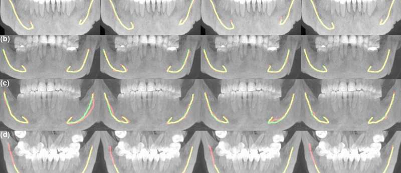
A dentist inserting a tooth implant must know the exact location of the nerve canal in the patient’s lower jaw to plan the size and position of the implant, along with the overall procedure. This requires X-ray images in which the dentist or radiologist manually specifies the location of the canal point by point. Studying and analyzing these images can be arduous and time-consuming.
Dental equipment manufacturer Planmeca, the Finnish Center for Artificial Intelligence (FCAI) and Tampere University Hospital (Tays) have joined forces to tackle the problem. The result is an AI-based model that locates the lower jaw nerve canal in 3D X-rays faster than a human and with better precision than other automated methods.
“The collaboration arose from the needs of experts practicing clinical work and from seeking ways to help their everyday work. A lot of time can be saved by using artificial intelligence in patient treatment planning,” says Vesa Varjonen, Vice President of Research and Technology at Planmeca.
The method is based on training deep neural networks with a mass of clinical data, comprised of three-dimensional images rendered with cone beam computed tomography (CBCT).
“Tampere University Hospital provided us with extensive and versatile clinical materials produced with several 3D-imaging devices. The data was divided at random and part of it used for training the neural networks and part of it isolated for testing and validating the designed method,” says Aalto University doctoral researcher Jaakko Sahlsten.
Artificial intelligence is an efficient and reliable tool
Nerves that control the motor functions of the jaw and facial senses run in the nerve canal of the lower jaw, the mandibular canal. In addition to implant placement, its location is crucial in wisdom teeth removal and jaw surgery. The location and route of the canal running inside the jawbone is unique to each person.
“One of the challenges in training the AI model was that the size of the mandibular canal in a 3D X-ray of the skull is very small compared to the data in the overall image. As a dataset, this type of training material is highly unbalanced,” Sahlsten notes.
Working together with Tays radiologists was key for harnessing the data into use when training artificial intelligence.
“When a huge amount of data is fed to the neural network and the location of the mandibular canal is marked in it, it learns to optimize its own internal parameters. The neural network resulting from this learning quickly finds the mandibular canal from the individual 3D data input,” Varjonen says.
Testing the neural network model with patient data isolated from the research materials demonstrated that the model managed to locate the mandibular canals with high precision: only 1–4% of the cases may be inaccurate.
“In clinical assessments, experts went through the results produced by the model and discovered that in 96% of the cases they were fully usable in clinical terms. We are highly confident that the model works well,” Sahlsten says.
Compared to humans, one of the advantages of artificial intelligence is that it always works with equal efficiency and speed. The AI model speeds up the discovery of the mandibular canal and supports radiologists’ and physicians’ decision-making. Final treatment decisions are always made by a health professional.
Publications verify model functionality
Planmeca is a Finnish family business and one of the world’s leading equipment manufacturers in health technology. Its products are exported to over 120 countries around the world. The company’s business is founded on 3D imaging devices for dental care and software that supports them. For Planmeca, collaboration with FCAI and Tays means significant new business potential.
“Digitality and AI used in imaging equipment are important for us. We will integrate the neural network model developed in this research into our imaging software. This will improve the usability and performance of our equipment,” Varjonen says.
The scientific publications produced in the collaboration are important for all project partners. Some of the results were published in Scientific Reports.
“Peer-reviewed publications are solid evidence of the functionality of the model. Deep learning has not previously been used in tasks of this type, which adds to the value of the publications. They also promote doctoral candidates’ thesis work,” Sahlsten says.
“The publications will be important for us when applying for a medical device approval for our software. They demonstrate that the software has been designed according to software development processes and scrutinized through all required phases,” Varjonen notes.
In addition to locating the lower jaw nerve canal, the collaborative project between Planmeca, FCAI and Tays also covered the development of a neural network model for orthognathic surgery, in which anomalies in the lower face area are corrected through surgical measures.
“The model helps to identify landmarks in the skull area for correcting malocclusion and planning jaw alignment surgery. The same patient data was also used for another AI application,” Varjonen says.
Going forward, artificial intelligence will have a lot to offer in health applications.
“I see artificial intelligence as a very powerful tool that physicians and other experts can use when making their first assessments or to get alternative opinions. The challenge with deep learning models is that we cannot give definite grounds as to why the model reaches a specific outcome. Further research is needed to increase the explainability and transparency of the models,” Sahlsten concludes.
More information:
Jorma Järnstedt et al, Comparison of deep learning segmentation and multigrader-annotated mandibular canals of multicenter CBCT scans, Scientific Reports (2022). DOI: 10.1038/s41598-022-20605-w
Journal information:
Scientific Reports
Source: Read Full Article


