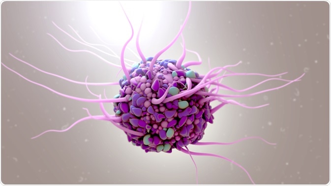
Recently, there has been an increase in the use of human cell lines for research purposes. However, this has also been accompanied by an increase in cross contamination. Short Tandem Repeat (STR) DNA profiling is one solution that has been proposed for the detection of cross-contamination and authentication of human cell lines.
 Design_Cells | Shutterstock
How often does cross-contamination occur?
Cross-contamination is a critical issue in cell culture, with studies suggesting that up to 16-35% of all experiments are affected. This impacts research as the recorded results may not accurately reflect the true data and replicating the experiment is difficult. Detecting contaminated cell lines is therfore imperative.
Detecting cross-contaminaton is particularly difficult for cells with similar morphologies or phenotypes. For example, the HeLa cell line, which was developed in 1951, now consists of more than 90 cell lines. In a recent study, different cell lines from breast, thyroid, adenoid, esophagus cancers were all found to be cross-contaminated. Thus, the use of methods that can identify cell lines has become crucial.
What is Short Tandem Repeat (STR) profiling?
Short Tandem Repeats refer to short DNA sequences that are identical between cells from the same cell lines, and lie successively in specific regions of the chromosomes. The size of short tandem repeats may range from two to thirteen nucleotides, and they are used to compare specific loci on DNA in different samples.
In 1985, Alec Jeffreys and his colleagues showed the presence of variable number tandem repeats distributed throughout the genome that can give rise to a characteristic signature of DNA. This can be used to identify particular cell lines. However, these fingerprints can be difficult to interpret. As a solution, short tandem repeats (STR) were used. STR consists of short sequences of size 1–6 base pairs.
STR profiling of human cell lines: Methodology
The short tandem repeat profiling can be carried out in any lab that has the capacity for molecular biology techniques. As soon as a cell line is received in a lab, all the information regarding its name, name of the donor, name of the original cell line, the tissue of origin, doubling period, its characteristics and functions are recorded.
For the development of the assay for adherent cells, the culture medium is first removed and discarded. The surface is then rinsed using PBS (which is subsequently discarded) and the cells are dissociated. This is followed by washing of the pellet and resuspension in PBS before counting the cells.
Storage conditions
The cells are spotted into an FTA card by suspending 200,000 cells in PBS. Then a 1.2 mm disk is punched into the center of the sample spot and a plunger is used to eject the disk into the reaction well plate.
The amplification process involves adding all the components of a PCR mixture in a sterile tube and vortexing for 10 seconds. Then 25 μl of PCR mix is added to the reaction tube that contains the FTA disk. Thermal cycling involves placing the plate in the thermal cycle and running the recommended protocol. The amplified samples can be stored at 4°C after completing this cycle.
Detection
Primers tagged with fluorescent markers recognize different loci in the PCR reaction. After the PCR is completed, the internal standards for size can be added to the reaction mixture and the DNA can be separated on the basis of size using capillary gel electrophoresis.
Size analysis is completed using the GeneMapper ID-X software, which compares the size of different DNA fragments with internal standards. The short tandem repeat genotyping is done by conversion of the amplicon sizes into alleles using the allelic ladder.
Authentication and data analysis
If the STR profile shows more than an 80% match, a cell line is considered authentic, while a match that is less than 56% is considered to be unrelated. In some cases, however, the match profile may be indeterminate if the similarity is between 56% and 80%. Usually, at least eight STR loci are required to establish the identity of a human cell line.
Sources
- Reid, Y. et al. (2013). Authentication of Human Cell Lines by STR DNA Profiling Analysis. Assay Guidance Manual. Eli Lilly & Company and the National Center for Advancing Translational Sciences. www.ncbi.nlm.nih.gov/books/NBK144066/
- Northwestern University. Human Cell Line Authentication through STR Profiling. www.cgm.northwestern.edu/cores/nuseq/other-services/cell-line.html
Further Reading
- All Cell Culture Content
- Common Problems in Cell Culture
- How to Generate Stable Cell Lines
- HEK293 Cells: Applications and Advantages
- What is Micropatterning?
Last Updated: May 2, 2019

Written by
Dr. Surat P
Dr. Surat graduated with a Ph.D. in Cell Biology and Mechanobiology from the Tata Institute of Fundamental Research (Mumbai, India) in 2016. Prior to her Ph.D., Surat studied for a Bachelor of Science (B.Sc.) degree in Zoology, during which she was the recipient of anIndian Academy of SciencesSummer Fellowship to study the proteins involved in AIDs. She produces feature articles on a wide range of topics, such as medical ethics, data manipulation, pseudoscience and superstition, education, and human evolution. She is passionate about science communication and writes articles covering all areas of the life sciences.
Source: Read Full Article


