In a recent study published in the Particle and Fibre Toxicology Journal, researchers explored the impact of exposure to ultrafine particles during pregnancy on the risk of influenza infection.
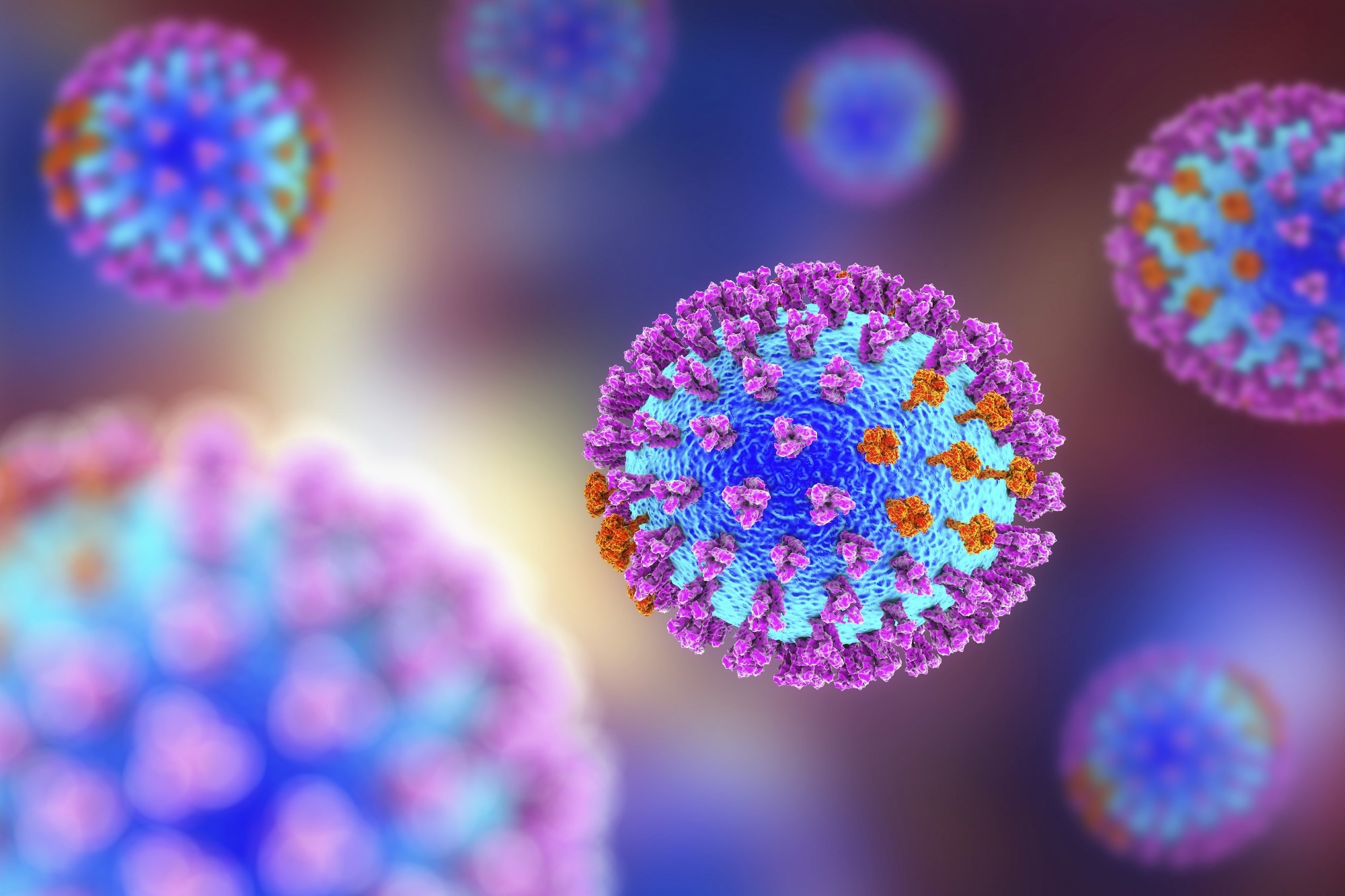
 Study: Maternal exposure to ultrafine particles enhances influenza infection during pregnancy. Image Credit: KaterynaKon/Shutterstock.com
Background
Identifying interactions between infectious agents and air pollution is crucial, particularly for safeguarding vulnerable populations.
The susceptibility of pregnant women to influenza and air pollution exposure is a matter of concern, but the relationship between the two is not fully understood.
Exposure to ultrafine particles (UFPs) in mothers can cause distinct immune responses in the lungs.
About the study
In the present study, researchers hypothesized that exposure to UFP during pregnancy could result in abnormal immune responses to influenza, which could increase the severity of the infection.
In the study, pregnant mice were exposed to either UFPs or filtered air (FA) equivalent to a 24-hour average of 25 µg/m3 during gestational days (GD) 0.5 to 13.5.
The study involved inoculating dams with either heat-inactivated (HI) control virus or live Influenza A/Puerto Rico/8/1934 (PR8) virus and evaluating them three days after infection.
The viral titer of PR8 was measured in the lung using a 50% tissue culture infectious dose (TCID50) assay and quantitative polymerase chain reaction (qPCR) to determine its infectivity.
A semi-quantitative scoring system was used to evaluate pulmonary histopathology three days after infection. The study examined T cell responses in maternal lung samples from different exposure groups, specifically T1, T2, T17, and CD8+cytotoxic T lymphocyte (CTL) lineages.
Results
Plug-positive female mice in all exposure cohorts had a pregnancy rate of at least 60%. UFP exposure did not impact pregnancy loss due to comparable pregnancy rates among the groups.
The FA-HI cohort did not show a significant difference in percent weight gain compared to the control mice housed in a different room. The percent weight gain estimated for FA- or UFP-exposed dams inoculated with PR8 showed significant differences, suggesting a strong infection.
Both infected groups impacted percent weight gain, suggesting a successful infection. No variations in the weight gain percentage were observed between UFP-HI and UFP-PR8 or between FA-PR8 and UFP-PR8.
The UFP-PR8 group had the highest expression in the qPCR examination of the influenza A segment, with a 3.7-fold increase compared to the FA-PR8 group.
The UFP exposure group showed more PR8 cytopathic effect (CPE) than the FA exposure group during TCID50 analysis. This was observed through cell lysis, excessive cell rounding, and multifocal cellular destruction.
Apart from one dam from the UFP-HI cohort, which displayed mild and focal inflammation, dams from the UFP-HI and FA-HI groups did not show significant inflammatory infiltration.
The FA-PR8 group showed mild to moderate inflammation, primarily in perivascular and peribronchial regions with lymphocytes, plasma cells, scattered neutrophils, and macrophages. The UFP-PR8 group showed only minimal aggregates of lymphocytes and no significant inflammation.
The UFP-PR8 cohort did not have inflammatory infiltrates, unlike the FA exposure group, indicating a suppression of the innate immune system.
The exposed and inoculated cohorts showed changes in their immune cell profiles, indicating potential lung immunosuppression. The UFP-PR8 group exhibited reduced levels of CD8+ cytotoxic lymphocytes.
The UFP-PR8 group showed a decrease in CD8+T cells, indicating early immune suppression in the adaptive arm. This is consistent with the reduced tissue pathology and high PR8 lung titer.
Five genes showed detectable expression. Sphk1 showed a moderate positive association with PR8 lung titer, while interferon regulatory factor 7 (Irf7), transforming growth factor (TGF-β), and interleukin-33 (IL-33) had little correlation.
The higher Sphk1 and Il-1β corresponded with a rise in viral titer. The UFP-PR8 group exhibited around five times higher levels of Sphk1 in comparison to the FA-PR8 group. Increased Sphk1 levels and activity are observed in severe cases of influenza infection, which can promote the replication of the virus.
The expression of Il-1β was significantly higher in the UFP-PR8 group compared to the FA-PR8 cohort.
Conclusion
The study findings highlighted various pieces of evidence that suggest a higher vulnerability to influenza infection due to exposure to gestational UFP.
These include reduced weight gain, increased viral titer, reduced lung inflammation and T-cell response, and changes in inflammatory mediators and pro-viral variables. The study's findings provide an initial basis for safeguarding pregnant women and managing UFPs through clinical and regulatory measures.
Vaccination for pregnant females is crucial in urban areas where influenza and UFPs are prevalent. Preventive actions are necessary to safeguard maternal health by limiting UFP exposure and promoting vaccination.
-
Drury, N. et al. (2023) "Maternal exposure to ultrafine particles enhances influenza infection during pregnancy", Particle and Fibre Toxicology, 20(1). doi: 10.1186/s12989-023-00521-1. https://particleandfibretoxicology.biomedcentral.com/articles/10.1186/s12989-023-00521-1
Posted in: Child Health News | Medical Science News | Medical Research News | Women's Health News | Disease/Infection News | Healthcare News
Tags: Air Pollution, Assay, Cell, Cell Lysis, Genes, Growth Factor, heat, Histopathology, Immune System, Immunosuppression, Inflammation, Influenza, Interferon, Interleukin, Lungs, Lymphocyte, Maternal Health, Neutrophils, Pathology, Pollution, Polymerase, Polymerase Chain Reaction, Pregnancy, T Lymphocyte, T-Cell, Tissue Culture, Toxicology, Virus

Written by
Bhavana Kunkalikar
Bhavana Kunkalikar is a medical writer based in Goa, India. Her academic background is in Pharmaceutical sciences and she holds a Bachelor's degree in Pharmacy. Her educational background allowed her to foster an interest in anatomical and physiological sciences. Her college project work based on ‘The manifestations and causes of sickle cell anemia’ formed the stepping stone to a life-long fascination with human pathophysiology.
Source: Read Full Article


