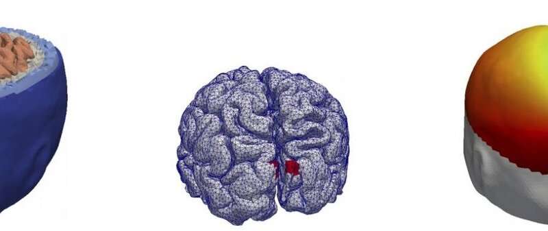
Skoltech researchers have proposed a fast and accurate numerical method of addressing the problem plaguing electroencephalography (EEG) studies that monitor the brain’s electrical activity—having to laboriously locate the source of EEG signal in the brain due to the low spatial resolution of this method. The new approach may help improve both medical and research applications of EEG. The paper was published in IEEE Transactions on Biomedical Engineering.
Suppose you want to study the properties and activity of a human brain without cracking open the brain owner’s skull (invasive research methods have their applications too, but those are understandably limited). You could put the brain, with its owner, into an MRI machine, and that’s how most of those trendy studies in the news are done. MRI can offer great spatial resolution in that you could locate brain activations quite accurately. But it is exasperatingly slow, capturing processes that take minutes when a human brain’s typical reaction times are in the span of tens and hundreds of milliseconds. Then there’s MEG, magnetoencephalography, which is very accurate and more attuned to the quick thinking of humans but requires extremely expensive equipment that needs to be cooled down with liquid helium and operated in a magnetically shielded room.
EEG, electroencephalography, however, is much simpler and easier to set up and use, and it provides a very good temporal resolution; that is why it is so widely used in healthcare and research. There’s just one catch, explains Mikhail Malovichko, a coauthor of the study: even a small active area of the cortex generates electrical potential on a large portion of the surface of the head, so an accurate localization of small active patches of the brain is a challenging mathematical task, the so-called inverse EEG problem.
To solve this problem, researchers normally use MRI scans to build a model of the subject’s head, place some candidate electric dipoles, essentially best guesses for where the signals might be coming from, and have a computer tinker with the model until its output fits the actual signal measured on the head. For this, the machine has to first solve many complementary forward problems: figure out what kinds of electrical activity these candidate dipoles would generate.
“This approach is universal. The preliminary solution of forward problems reduces the inverse EEG problem to a small system of linear equations, which is of the same type regardless of the position of candidate dipoles and the numerical method used to solve the forward problem. But if one needs to consider each subject’s anatomical features, then the forward problem has to be solved by the finite element method, a very resource-intensive numerical procedure,” says Nikolay Koshev, another coauthor of the study.
That takes quite a lot of time, so Malovichko and his colleagues from the Skoltech Center for Data-Intensive Science and Engineering (CDISE) have proposed to approach this challenge in a different way. Their solution for the inverse EEG problem directly “backpropagates” measured signals from the skin inside the head down to the cortex. This requires reframing the whole task as a Cauchy problem, a type of mathematical problem that is known to be unstable for EEG: that means even slight deviations in the input, for instance, from unavoidable measurement errors, can significantly skew the result. Yet recent research has brought new approaches to tackling these unstable problems efficiently, and the scientists used them in their research.
“In essence, instead of treating each candidate electric dipole separately and having to solve the forward problem first for each of them, the algorithm now has to solve just one inverse problem, which is, however, of a rather peculiar kind. This helps speed up the processing of EEG data and increases accuracy for source localization; in addition, the algorithm explicitly incorporates the information on how the brain surface is shaped,” Mikhail Malovichko says.
Source: Read Full Article


