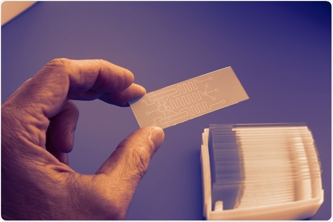
A protein microarray consists of the immobilization of proteins in organized rows and columns upon a support structure. The structure of the protein microarray allows for high-throughput analysis of protein functions and interactions.

Credit: science photo/ Shutterstock.com
An early use of the technology was for the detection of protein binding abilities. The interaction between two proteins is investigated by placing one protein on the array in multiple samples. Each sample is then probed with a fluorescently labelled protein, with interactions identified by a fluorescent sample spot. Various protein interactions have been investigated, including antibody-antigen pairing for use in clinical diagnostics.
Microarrays permit the production of high sample quantities at a fast rate with inexpensive production costs. The technology has increased the ease of handling, as well as the level of reproducibility. While there are clear advantages to protein microarrays, their application is not without challenges.
Challenges to developing protein microarrays
Protein microarrays were developed from the more commonly employed DNA microarray. The requirements of protein microarrays are nevertheless more complex and have required material customized from classic DNA microarrays to make them suitable.
Proteins are vulnerable to denaturation through changes in pH and temperature. Furthermore, while DNA can simply be immobilized through an electrostatic interaction, proteins have a more variable surface charge, which means the material used for DNA microarrays has to be adapted.
For antibody microarrays, the antibodies must remain active in order to bind to the analyte on the surface. This requires an immobilizing material that preserves the active stage of the antibody for long periods of time in storage.
One method of arraying functionally active proteins is immobilization through covalent bonding to the smooth surface of the glass slide. Types of immobilizing agents include aluminum, gold and hydrophilic polymers, which are all applied as coating to the support surface of the array.
Challenges to quantifying protein concentrations
As protein microarrays provide the possibility of analyzing hundreds of interactions between antibodies and antigens on one array, the challenge of quantifying proteins concentrations has been introduced. There can be a six fold variation of within-cell protein concentrations, which means that detection methods need to be able to quantify protein concentrations with different orders of magnitude.
An additional problem of false negatives may also be produced from the basic investigation of protein interactions on an array. The conjugation of proteins on a microarray can change the folding structure of the protein, destroying the antibody-antigen pairing and thus leading to a false negative result.
Overcoming challenges in both label-based and label free protein microarrays
The challenge to refine protein microarrays for utilization towards the level of DNA microarrays is currently under way. There are now two strategies for protein microarray detection: label-based and label free.
Label-based microarrays use the traditional method of labelling probes through materials such as fluorescent dyes or radioisotopes. In contrast, label free microarrays measure an inherent property of the probe sample, such as its mass. Though label-based protein microarrays have simple instrument requirements and use widely available reagents, the labelling process can alter the surface characteristics of the molecule in question.
The development of a label-free approach avoids interference to the molecule within the probe. The application of surface plasmon resonance (SPR) is an example of this technique. Proteins are immobilized on the microarray with a thin, gold coating placed on the surface structure. The unlabeled protein probe is then added, and a change in the angle of the reflection of light caused by the interaction between the two proteins is used for detection.
In comparison to label-based methods, the sensitivity and specificity of unlabeled methods such as SPR are lower. However, their potential for refinement (along with the ability to provide quantitative information about protein binding kinetics) means that they will be increasingly utilized in the future.
Reviewed by: Dr Tomislav Meštrović, MD, PhD
Sources:
- http://www.sciencedirect.com/science/article/pii/S0021967303007696
- https://www.ncbi.nlm.nih.gov/pubmed/15239896
- http://europepmc.org/abstract/med/15782547
- https://www.ncbi.nlm.nih.gov/pubmed/19953541
Further Reading
- All Microarray Content
- Types of Microarray
- Microarray Applications
- Protein and Peptide Microarray
- History of Microarrays
Last Updated: May 24, 2019

Written by
Shelley Farrar Stoakes
Shelley has a Master's degree in Human Evolution from the University of Liverpool and is currently working on her Ph.D, researching comparative primate and human skeletal anatomy. She is passionate about science communication with a particular focus on reporting the latest science news and discoveries to a broad audience. Outside of her research and science writing, Shelley enjoys reading, discovering new bands in her home city and going on long dog walks.
Source: Read Full Article


