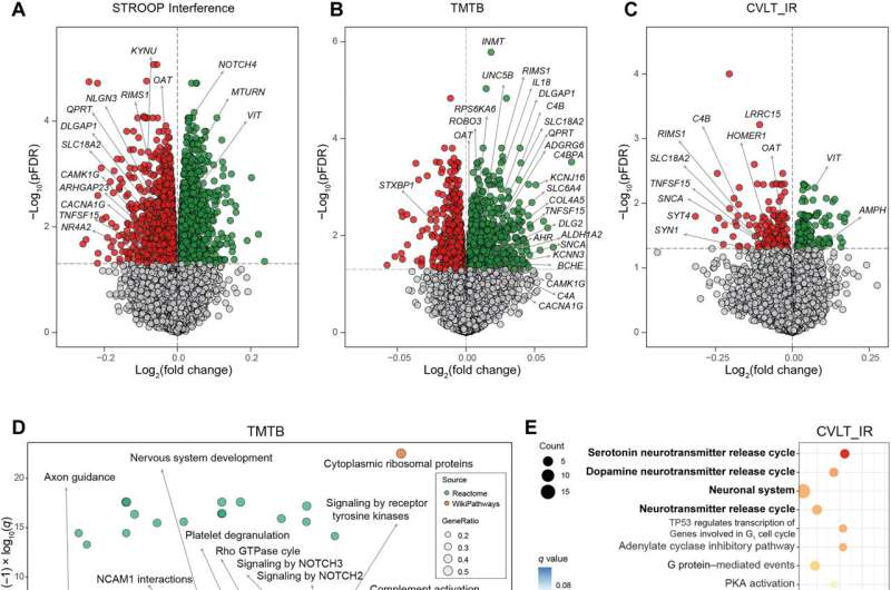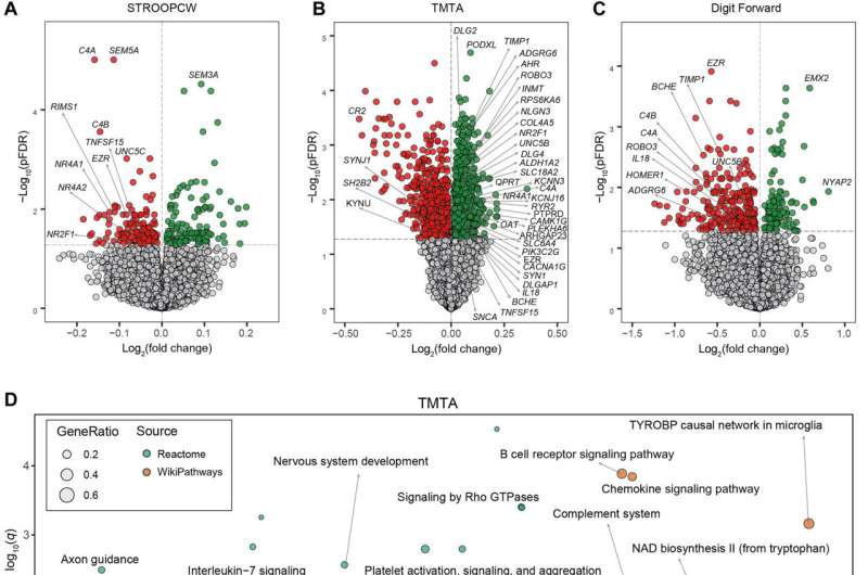
In mice, cognitive decline is influenced by obesity due to the potential association of an adipose-brain axis. To examine this proposal, Núria Olivera-Canellas, and a team of researchers in the departments of medicine and diabetes at the University of Girona, Spain, identified 188 genes by using RNA sequencing of adipose tissue in three patient cohorts associated with cognitive performance. The genes had associations with synaptic function, anti-inflammatory signaling, vitamin metabolism, phosphatidylinositol metabolism and the complement cascade.
The team translated these findings into the plasma metabolome, and linked the circulating blood expression levels of the genes with several cognitive domains across a cohort of 816 participants. They showed how the targeted misexpression of the candidate gene orthologs in the Drosophila fat body significantly altered memory and learning in the flies.
In addition, the downregulation of a neurotransmitter release cycle-associated gene improved the cognitive abilities of both Drosophila flies and mice. The results showed hitherto unidentified connections between the adipose tissue transcriptome and brain function to gain unprecedented insight into the diagnostic and therapeutic targets in adipose tissue. The study is published in Science Advances.
The impact of obesity on the brain
Peripheral tissues can regulate brain function to shape different cognitive domains. Biologists have conducted studies in rats to reveal how restoring glucose metabolism of myeloid cells reversed cognitive decline in aging. Additionally, the selective ablation of gastrointestinal vagal afferent nerves impaired the hippocampus-dependent episodic and spatial memory in rats. These studies highlighted the liver to play a major role in determining feeding behavior in mice.
While obesity is linked to cognitive impaired risks, the increased body-mass index during middle-age predicts decreased cognitive abilities, which mainly include memory, executive functioning, and learning. However, precise mechanisms underlying the process remain largely unknown.
The brain can regulate the performance of adipose tissue. Olivera-Canellas and team therefore studied the mechanisms underlying directional interactions between adipose tissue gene expression and cognitive function in this work. They used RNA-sequencing analysis of adipose tissue to show genes associated with diverse cognitive domains. The scientists also explored the expression of several genes in peripheral blood mononuclear cells in association with cognitive functions in humans.
Genes involved with obesity and cognition
The researchers gained insight into the fat-brain axis by performing transcriptomics after RNA-sequencing the visceral adipose tissue obtained from 17 individuals with a severe obesity index between the ages of 28 to 60. They conducted different cognitive tests to assess the functional performance of memory, executive function and attention. The outcomes did not show significant associations with physical activity, macronutrients, glycemia or alcohol intake.
The team noted significant associations with gene transcripts for all cognitive tests, which included variants across diverse cognitive domains. Proteins encoded by these genes showed functions in the central nervous system, although their expression in adipose tissue in association with cognition is unprecedented.
Of the identified genes, the NUDT2 gene is associated with intellectual disability, AMPH encodes for a protein that is altered in the cerebrospinal fluid of patients with mild cognitive impairment and Alzheimer’s. OAT is another gene that plays a significant cognitive role, while KYNU and INMT genes are involved in tryptophan metabolism. Obese subjects had impaired memory and altered tryptophan metabolism.
To accurately interpret and identify the adipose tissue pathway associated with cognitive function, Olivera-Canellas and team mapped significant genes to the Reactome and WikiPathways databases included in ConsensusPathDB. The team observed an overrepresentation of pathways with an important role in the central nervous system associated with inhibitory control, working memory, immediate memory and attention. They also noted an overrepresentation of pathways associated with inflammation-linked cognitive domains, as well as altered notch signaling and complement activation associations with working memory.

Adipose tissue gene expression is associated with several cognitive functions later in life
Olivera-Canellas and colleagues validated the outcomes by performing RNA-sequencing of visceral adipose tissue and subcutaneous adipose tissue across 22 subjects with severe obesity between the ages of 23 to 57 years. The cohort of patients underwent the same battery of cognitive tests, which were performed two to three years after adipose tissue sample collection. As before, the scientists did not observe a significant association of physical activity, diet, alcohol intake and glucose or hemoglobin levels. They noted a significant gene association with the cognitive test.
In total, the team noted axon guidance, nervous system development, neuronal system and tryptophan pathways associated with attention, while the complement system activated in visceral adipose tissue to indicate a strong association with performance in several cognitive domains. The subcutaneous adipose tissue pathways were associated with synaptic transmission, neuronal system, and one-carbon metabolism relative to long-term and short-term memory.
Further experiments on cognition and adipose tissue-related genes in the adipose-brain axis
The scientists further noted the association of adipose tissue genes linked to cognitive function in the neuronal system and during synaptic formation of the pathways. Those overrepresented genes showed key roles in synaptic function and inflammation.
The team conducted additional experiments to link adipose tissue to cognition alongside its associations with inflammation. The outcomes highlighted morbid obesity to influence the association between adipose tissue genes and cognition, while linking the blood expression levels of genes to cognitive performance.
The researchers used fruit flies (Drosophila melanogaster) to experiment the causal effect of the adipose tissue-related expression of candidate genes to promote cognition. Added analysis included cognition tests in mice adipose tissue or Drosophila fat body to understand the cluster of genes involved in the release of neurotransmitters.
Outlook
In this way, Núria Olivera-Canellas and colleagues showed how the brain regulated adipose tissue performance while the reverse is also plausible. The expression of genes related to axon guidance, nervous system development, neuronal system, tryptophan and inflammation were all linked to cognition across three different cohorts.
Together with added studies in animal models, the researchers suggest a systemic developmental process that changed in diverse tissues and cells as reflected in neuronal networks. The outcomes provide useful biomarkers to predict cognitive decline and monitor response to treatment.
More information:
Núria Oliveras-Cañellas et al, Adipose tissue coregulates cognitive function, Science Advances (2023). DOI: 10.1126/sciadv.adg4017
Paras S. Minhas et al, Restoring metabolism of myeloid cells reverses cognitive decline in ageing, Nature (2021). DOI: 10.1038/s41586-020-03160-0
Journal information:
Science Advances
,
Nature
Source: Read Full Article


