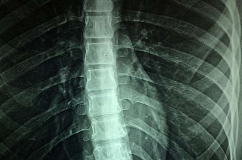
A team of researchers at The Ohio State University Wexner Medical Center and College of Medicine have developed the first mouse model capable of reproducing the ischemic spinal cord injury (“ISCI”) and immediate paralysis that sometimes result from a common procedure used to treat thoracic aortic aneurysm.
Study findings are published online in the journal Anesthesiology.
The procedure, known as “TEVAR” (thoracic endovascular aortic repair), can be a life-saving treatment that repairs aortic aneurysms. However, patients undergoing TEVAR are at risk of developing paralysis caused by ISCI as a result of disruptions to spinal cord blood flow during the procedure.
The new mouse model will enable researchers to study the molecular mechanism of paralysis caused by TEVAR. Understanding these mechanisms is a crucial step toward developing neuroprotective drugs and therapeutics to prevent or treat TEVAR-induced ISCI and paralysis.
TEVAR is used to treat an aneurysm, a weak balloon-like bulge, in the upper part of the aorta, the essential blood vessel that supplies blood to the whole body. If the aneurysm bursts, it is a medical emergency that can be deadly.
During the TEVAR procedure, a stent graft—a metal tube covered in fabric—is placed into the aorta to reinforce the aneurysm and prevent it from bursting. Ischemic spinal cord injury and paralysis occur when the stent graft blocks blood flow to the arteries that branch off from the aorta, reducing blood flow to the spinal cord (a condition known as “hypoperfusion”). Patients who develop paralysis after TEVAR have a significantly lower survival rate compared to non-paralyzed patients.
“The importance of this model cannot be overstated,” said Dr. Hesham Kelani, a former post-doctoral research fellow at Ohio State Wexner Medical Center, lead author and inventor of the new mouse model.
According to the Society for Vascular Surgery, 215,000 people in the United States are diagnosed with an aortic aneurysm every year. Every individual who undergoes TEVAR is vulnerable to spinal cord injury and possible paralysis. Approximately 3.7% of patients who undergo TEVAR suffer this devastating complication.
“This is the first and only mouse model that represents this type of injury in humans,” said Kelani, who led the team of Ohio State researchers. “Despite the frequency of post-TEVAR paralysis, in the 30 years since the first TEVAR procedure, scientists have never had a mouse model mimicking TEVAR-induced ISCI and have therefore been unable to study the mechanisms and pathophysiology of spinal cord hypoperfusion caused by TEVAR.”
That is, until now.
To create the mouse model, Kelani induced spinal cord hypoperfusion and ISCI in adult mice by ligating (or “tying”) five pairs of the arteries branching off from the aorta (known as intercostal arteries) located in the chest of the mice. The resultant mobility and histopathological changes mimicked those experienced by human patients after TEVAR.
As with human patients, paralysis was immediate, and mildly paralyzed mice showed progressive improvement of motor function over time, while severely paralyzed mice demonstrated permanent loss of motor function.
“This study also pioneers the use of a laser Doppler in vivo on a mouse spinal cord and intercostal arteries to document the extent of arterial ligation and the resulting spinal cord hypoperfusion,” Kelani said.
The Ohio State team included researchers from Anesthesiology, Veterinary Biosciences, Emergency Medicine, Physiology and Cell Biology, Biomedical Informatics and Neuroscience, the School of Health and Rehabilitation Sciences and the Center for Electron Microscopy and Analysis.
The work was performed under the supervision of D. Michele Basso, associate director of Ohio State’s School of Health and Rehabilitation Sciences, and creator of the Basso Mouse Scale for Locomotion. This research is the latest example of Ohio State’s leadership in the study of ischemic spinal cord injury from aortic aneurysm repair. Twelve years ago, a research team led by Dr. Hamdy Elsayed-Awad, professor of anesthesiology and senior author, created a successful mouse model mimicking ISCI caused by open surgical repair of an aortic aneurysm.
“This collaborative work takes us much closer to finding treatments for ischemic spinal cord injury by now being able to model the most up-to-date surgical and therapeutic approaches for aortic aneurysms. Our continued work in this important unexplored area may help prevent this debilitating paralysis in the future,” Basso said.
More information:
Hesham Kelani et al, A Mouse Model of Spinal Cord Hypoperfusion with Immediate Paralysis Caused by Endovascular Repair of Thoracic Aortic Aneurysm, Anesthesiology (2023). DOI: 10.1097/ALN.0000000000004515
Journal information:
Anesthesiology
Source: Read Full Article


