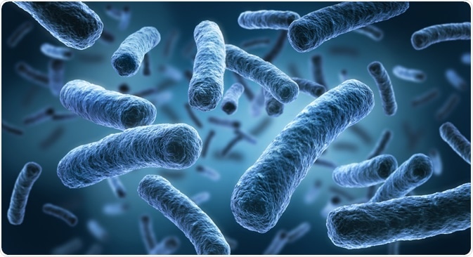
Flow cytometry has been a vital tool in microbiology and immunology for its ability to provide quantitative data at the single-cell level. Thanks to recent developments that have led to improved resolution, flow cytometry can now be applied to the detection and analysis of single-celled microbes.

Image Credit: peterschreiber.media/Shutterstock.com
How can flow cytometry be beneficial in microbiology?
Determining the presence and size of microbial populations in cell populations is critical for several industries, including research laboratory settings and food production. For example, while the presence of certain yeasts can be beneficial in beverage production, the presence of other non-desired microbes needs to be monitored.
Most traditional methods of microbial detection and characterization require isolation of the target organism, with subsequent culture growth which can take 40 to 72 hours. Flow cytometry can allow for quick detecting, counting, and characterization of microbes by labeling them with fluorescent dyes.
This method is quicker than traditional methods and allows for the analysis of more cells. Furthermore, this means that slow-growing microbes, such as certain fungi, can be studied more efficiently.
Further developments, including improvements in the resolution that allows for analysis of smaller individual cells and increased dye availability for bacterial cells, are constantly expanding the field of flow cytometry for use on microbes. The chief benefit of flow cytometry for microbes is that it can be applied to cell populations of mixed origin.
How flow cytometry works
Different types of flow cytometers can be used for different purposes, such as measuring cellular parameters or sorting cell subsets. Cells are passed sequentially through the instrument, where a light source illuminates the sample.
The light and scatter are absorbed by a photodetector, which converts the light into photocurrent that can be stored and analyzed quantitatively.
If fluorescence is not used, scatter light can still be collected to understand more basic parameters such as cell size and complexity (number of organelles inside the cell). When fluorescence is used, the laser excites fluorophores that have been tagged to the sample and these photons are absorbed by the photodetector.
The type of dye can influence the application the flow cytometry is used since it targets different substrates. For example, when a dye such as Texas Red targets proteins, flow cytometry can be used for microbe detection. However, if a dye targeting DNA or RNA, such as SYTOX Green, is used, flow cytometry can be used for DNA quantification.
Applications
Flow cytometry on microbes has several important applications, including in research, clinical, and industrial settings. Most of these are relevant for the detection and analysis of microbes in environments where other cells or microbes may also be present.
As mentioned, one of the foremost benefits of flow cytometry is its ability to detect microbes against a complicated background of other cells and/or other microbes. This has made flow cytometry particularly useful for complex screening, such as those involved in quality control of food and beverage samples.
Similarly, flow cytometry can be used to determine microbial content in pharmaceutical laboratories where water, raw materials, and products are tested.
In settings where samples from the environment are taken, it can be possible to encounter previously unidentified microbes. In these cases, traditional means of analysis would struggle, as these would require culture growth and culture requirements are not known. Flow cytometry can target these unidentified microbes by using fluorescently labeled probes that target the 16S ribosomal RNA of microbes.
Clinical settings are also greatly benefitting from the applications of flow cytometry. Diseases caused by microbes are overwhelmingly common, and flow cytometry can be crucial in detecting these in body samples.
Furthermore, studies on DNA quantification using flow cytometry with certain fluorescent dyes can also be used to study the microbe’s cell cycle, and thus have important implications for the viability and spread of an infectious microbe.
Similarly, flow cytometry has successfully been used to study the interaction between pathogens and phagocytic cells. This has been possible due to fluorescent labels that can detect oxidative bursts that occur because of the phagocytic process. So far, this has been successfully applied to yeast strains, Salmonella, and Escherichia coli, among others.
In more extreme cases, flow cytometry has also been suggested as a possible screening mechanism to prevent biological terrorism or warfare. Highly lethal microbes, such as Bacillus anthracis which causes anthrax, it is the spores rather than microbial cells that cause deleterious effects.
However, flow cytometry has been successfully used to detect them, since fluorescent stains generally do not penetrate the spore coat and can thus be distinguished from other microbes.

Image Credit: Alexander Raths/Shutterstock.com
Sources
- Thermofisher Scientific. 2020. Flow Cytometric Detection of Bacteria in Research and Industrial Samples. [online] Available at: <https://www.thermofisher.com/au/en/home/references/newsletters-and-journals/bioprobes-journal-of-cell-biology-applications/bioprobes-79/bacteria-detection-research-industry-flow-cytometry.html>
- Alvarez-Barrientos, A., Arroyo, J., Canton, R., Nombela, C., and Sanchez-Perez, M., 2000. Applications of Flow Cytometry to Clinical Microbiology. Clinical Microbiology Reviews, 13(2), pp.167-195.
- Davey, H., 2002. Flow Cytometric for the Detection of Microorganisms. Methods in Cell Science, 24, pp.91-97.
Further Reading
- All Flow Cytometry Content
- Flow Cytometry Methodology, Uses, and Data Analysis
- Flow Cytometry Techniques used in Medicine and Research
- Flow Cytometry History
- Fluorescence-Activated Cell Sorting
Last Updated: Aug 3, 2020

Written by
Sara Ryding
Sara is a passionate life sciences writer who specializes in zoology and ornithology. She is currently completing a Ph.D. at Deakin University in Australia which focuses on how the beaks of birds change with global warming.
Source: Read Full Article


