Reflection-contrast microscopy is a type of light microscopy that can be used to analyze single cells, biopsies, and other small objects. In contrary to most light microscopy techniques, reflection-contrast microscopy requires extremely thin sections, which are normally used in electron microscopy.
This microscopy technique is, therefore, said to bridge the gap between light microscopy and electron microscopy and is considered to be a very important analytical tool.
Skip to:
- How does reflection-contrast microscopy differ from conventional light microscopy?
- How is high contrast achieved?
- Reflection-contrast microscopy and electron microscopy
- Applications of reflection-contrast microscopy
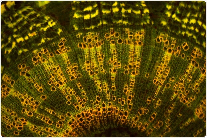 D. Kucharski K. Kucharska | Shutterstock
How does reflection-contrast microscopy differ from conventional light microscopy?
One of the most simple light microscopy techniques is bright field microscopy, and reflection-contrast microscopy has several important differences to this.
In bright field microscopy, transmitted light that has been shone through a sample is detected to form an image, whereas in reflection-contrast microscopy, images are formed from an incident light source reflecting off of a sample. This causes large differences in the output image produced.
Reflection-contrast microscopy has both a higher sensitivity and gives a higher definition than bright field microscopy. As is apparent in the title, reflection contrast microscopy provides a very high resolution and contrast. This means it is very easy to distinguish different parts of cells and their histology.
How is high contrast achieved?
There are two techniques used in reflection-contrast microscopy which greatly increase the contrast of the image; these are the central stop system and the antiflex principle.
The central stop system blocks the central beam of light from reflecting off the underside of the glass slide which the sample is mounted upon. This would normally cause blurring of the image, and decrease the contrast of the image.
The antiflex principle is put into place to contain unnecessary light rays, and limit the image to only be produced by reflected light. Using a combination of different plates and reflectors, only the light that has been reflected off the sample can pass through to form the image, whereas other stray beams of light are stopped.
Reflection-contrast microscopy and electron microscopy
The images produced by reflection-contrast microscopy can also look very similar to the images produced by low magnification transmission electron microscopy (TEM). Unlike light microscopy, TEM uses a thin beam of electrons which is partially transmitted through the sample to produce an image. This requires very specific conditions, which leads to TEM being a very expensive technique.
reflection-contrast microscopy can, therefore, provide a cheaper and easier alternative to low magnification transmission electron microscopy, as the contrast produced by reflection-contrast microscopy gives a very similar quality of the image.
Another advantage of reflection-contrast microscopy over electron microscopy is that reflection-contrast microscopy can be used to study living cells. TEM, on the other hand, needs to be performed within a vacuum, so cells cannot survive.
These two methods share similar preparatory steps, as they both use ultra-thin sections, so the same sample can be used in both techniques. At the very least, reflection-contrast microscopycan provide a simpler initial step for analysis of a sample, before further study using the high magnification power of TEM.
Applications of reflection-contrast microscopy
Reflection-contrast microscopy can be used for many common applications of microscopy to provide higher contrast and thus, an image of a higher quality.
Potential applications include in situ hybridization where specific labeled DNA or RNA can be located in a cell, detection of foreign material within a cell, detection of labeled trace molecules in a sample, and immunolocalization in cells and tissues.
As with many microscopy techniques, reflection-contrast microscopy can be adapted for many different research scenarios. The ability of the method to form a high contrast and sensitivity while still having a basic principle opens up many future applications for small scale research when electron microscopy equipment is not readily available.
Sources
- Taatjes, D. J. & Mossman, B. T. (2006). Cell Imaging Techniques. Humana Press. doi.org/10.1007/978-1-59259-993-6.
- Hayley Anderson. Transmission Electron Microscope. microscopemaster.com.
- Ploem, J. S. & Prins, F. A. (2017). Reflection-Contrast Microscopy – Review. Royal Microscopical Society. doi.org/10.22443/rms.inf.1.152.
Further Reading
- All Microscopy Content
- Advances in Fluorescence Microscopy
- Applications in Light Microscopy
- Electron Microscopy: An Overview
- Brief History of Microscopy
Last Updated: May 22, 2019
Written by
Jack Davis
Jack is a freelance scientific writer with research experience in molecular biology, genetics, human anatomy and physiology, and advanced analytical chemistry. He is also highly knowledgeable about DNA technology, drug analysis, human disease, and biotechnology.
Source: Read Full Article


