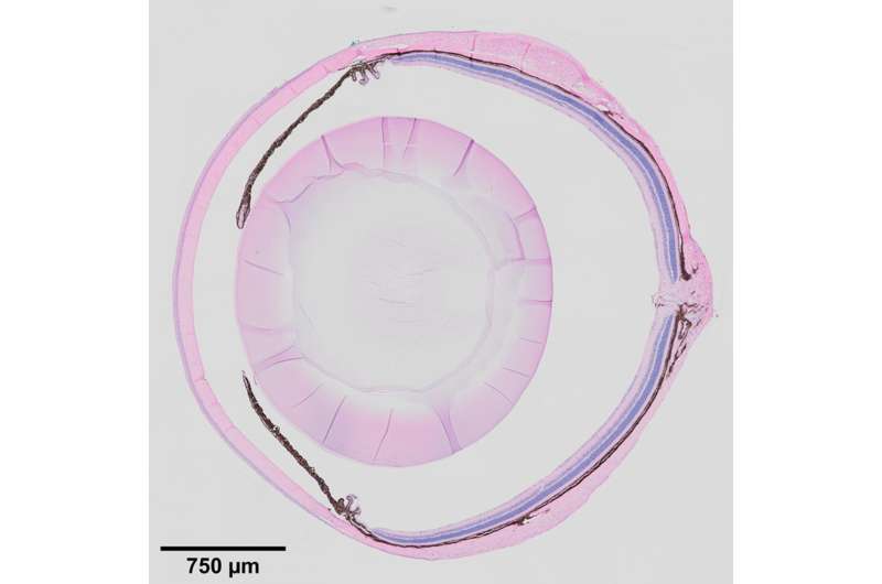
What if the degenerative eye conditions that lead to glaucoma, corneal dystrophy, and cataracts could be detected and treated before vision is impaired? Recent findings from the lab of Investigator Ting Xie, Ph.D., at the Stowers Institute for Medical Research point to the ciliary body as a key to unlocking this possibility.
Previous work from the lab showed that when mouse stem cells were differentiated into light-sensing photoreceptor cells in vitro, and then transplanted back into mice with a degenerative condition of the retina, they could partially restore vision. However, the transplanted photoreceptors only lasted three to four months.
“You cannot cure the condition in a diseased eye if you don’t know what causes the disease,” says Xie. “This has been a major hurdle for stem cell therapy in treating degenerative diseases.”
To this end, Xie’s group began to study the eye tissue microenvironment, specifically a specialized tissue in the eye called the ciliary body. Located at the posterior edge of the iris, it is known to maintain ocular pressure by secreting aqueous humor, the clear fluid between the lens and the cornea. It has a similar function in mice and in humans, and defects in the ciliary body manifest in similar ways in the mouse and human eye.
“People think the ciliary body is boring,” says Xie. This might be because the ciliary body was once thought to have a reserve of retinal stem cells, Xie explains, which turned out not to be true. However, its role in eye biology turns out to be quite broad, and “without a functioning ciliary body, the eye degenerates,” Xie adds.
When the Notch signaling pathway—an important cell signaling system found across the animal kingdom—is defective in the ciliary bodies of newly born mice, they fail to develop folds, and secretions decrease, leading to shrunken vitreous bodies. In adult mice, defects in Notch signaling cause low eye pressure, a shrunken vitreous, and eye degeneration. Inactivation of the downstream transcription factor RBPJ in the ciliary body also leads to the same effects. Before now, the underlying molecular mechanism for this outcome was unclear.
In a paper published in Cell Reports on January 12, 2021, first author Ji Pang, a visiting Ph.D. student from Shanghai Jiao Tong University, China, and others describe a signaling pathway wherein Notch and Nectin proteins in the ciliary body function in the development and maintenance of eye tissue and structure.
In this report, the researchers describe the roles of adhesion protein Nectin1 and gap junction protein Connexin43 in the ciliary body of mice. They found that Notch2/3-Rbpj signaling in the outer ciliary epithelium controls the expression of Nectin1, which works with Nectin3 in the inner ciliary epithelium to keep the two tissue layers together, which promotes proper folding of the ciliary body. They found that Notch signaling also maintains the expression of Connexin43 in the outer ciliary epithelium, while Nectin1 localizes and stabilizes Connexin43 on the lateral surface, which maintains the vitreous body and intraocular pressure.
Lastly, the researchers found that in addition to maintaining ocular pressure and directing ciliary body morphogenesis, Notch2/3-Rbpj signaling in the inner ciliary epithelium also regulates the secretion of various proteins such as Opticin and collagens into the vitreous body, providing nutritive support for the cornea, the lens, and the retina.
Source: Read Full Article


