The colorful pictures of the whole mouse brain at different ages are the first of their kind and a pivotal step forward in understanding behavior, scientists say.
Findings—published in the journal Science – could shed light on learning disability and dementia and help to reveal how memories are affected by age.
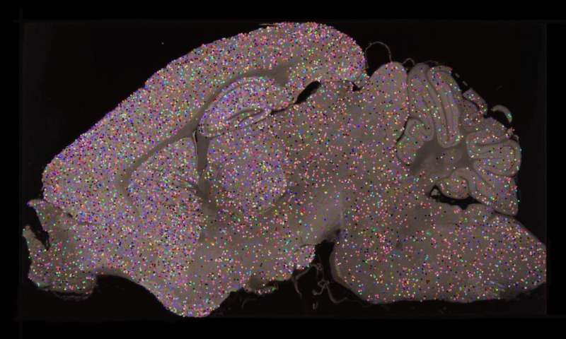
The images are of synapses—vital connections that carry electrical and chemical messages between brain cells. Synapses store memories and synapse damage is linked to more than 130 brain diseases.
Researchers based at the University of Edinburgh color-coded the different types of molecules to highlight the range of synapses in mouse brains from birth to old age.
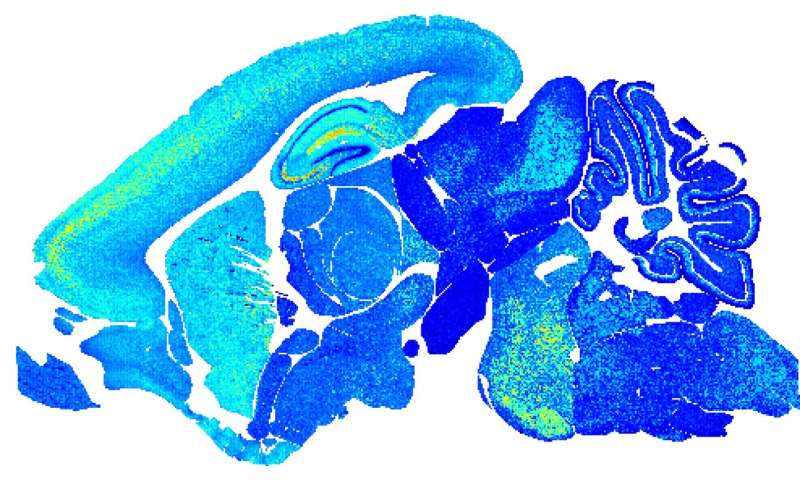
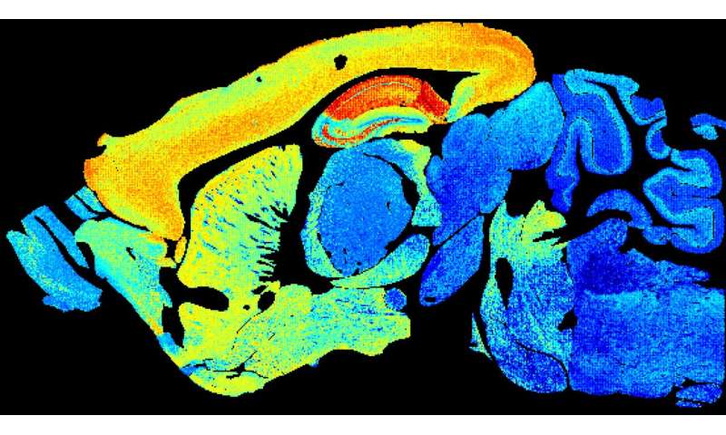
They discovered that the number and molecular makeup of synapses shifts with age in different parts of the brain. This happens at three main phases—childhood, middle and elderly age.
Synapse type shifts with age in patterns unique to areas of the brain, blossoming into a diverse array in midlife.
Images from middle-aged brains burst with color, illustrating a wide variety of synapses. Both very young and very old brain show less synapses and less complexity.
Researchers say these changes give insights into why genes cause synapse damage at set ages and in set brain areas.
The findings could shed light on why we are more likely to develop brain conditions at certain ages, helping to explain why schizophrenia often starts in adolescence, or why dementia affects older adults.
Lead researcher, Professor Seth Grant of the Centre for Clinical Brain Sciences at the University of Edinburgh, said: “The brain is the most complex thing we know of and understanding it at this level of detail is a momentous step forward.
Source: Read Full Article


